
Advertisement
-
Published Date
February 29, 2020This ad was originally published on this date and may contain an offer that is no longer valid. To learn more about this business and its most recent offers, click here.
Ad Text
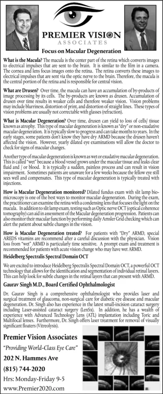
PREMIER VISION ASSOCIATES Focus on Macular Degeneration What is the Macula? The macula is the center part of the retina which converts images to electrical impulses that are sent to the brain. It is similar to the film in a camera. The cornea and lens focus images onto the retina. The retina converts these images to electrical impulses that are sent via the optic nerve to the brain. Therefore, the macula is the central portion of the retina and is responsible for central vision. What are Drusen? Over time, the macula can have an accumulation of by-products of image processing by its cells. The by-products are known as drusen. Accumulation of drusen over time results in weaker cells and therefore weaker vision. Vision problems may include blurriness, distortion of print, and distortion of straight lines. These types of vision problems are usually not correctable with glasses (refraction). What is Macular Degeneration? Over time, drusen can yield to loss of cells tissue known as atrophy. This type of macular degeneration is known as "dry" or non-exudative macular degeneration. It is typically slow to progress and can take months to years, In the early stages, some patients don't know they have dry ARMD because the drusen haven't affected the vision. However, yearly dilated cye examinations will allow the doctor to check for signs of macular changes. Another type of macular degeneration is knownas wet orexudative macular degeneration. This is called "wet" because a blood vessel grows under the macular tissue and leaks clear fluid and / or blood. It is typically a sudden, painless process and can result in vision impairment. Sometimes patients are unaware for a few weeks because the fellow eye still sees well and compensates. This type of macular degeneration is typically treated with injections. How is Macular Degeneration monitored? Dilated fundus exam with slit lamp bio- microscopy is one of the best ways to monitor macular degeneration. During the exam, the practitioner can examine the retina with a condensing lens that focuses the light on the macula. In addition to slit lampexam, testing such as Optic nerve OCT (optical coherence tomography) can aid in assessment of the Macular degeneration progression. Patients can also monitor their macular function by performing daily Amsler Grid checking which can alert the patient about subtle changes in the vision. How is Macular Degeneration treated? For patients with "Dry" ARMD, special AREDS vitamins are recommend after a careful discussion with the physician. Vision loss from "wet" ARMD is particularly time sensitive. A prompt exam and treatment is recommended for patients with acute vision change who may have wet ARMD. Heidelberg Spectralis Spectral Domain OCT We are excited to introduce Heidelberg Spectralis Spectral Domain OCT, a powerful OCT technology that allows for the identification and segmentation of individual retinal layers. This can help look for subtle changes in the retinal layers that can present with ARMD. Gaurav Singh M.D., Board Certified Ophthalmologist Dr. Gaurav Singh is a comprehensive ophthalmologist who provides laser and surgical treatment of glaucoma, non-surgical care for diabetic eye disease and macular degeneration. Dr. Singh also has experience in the latest small-incision cataract surgery including Laser-assisted cataract surgery (LenSx). In addition, he has a wealth of experience with Advanced Technology Lens (ATL) implantation including Toric and Multifocal lenses. Furthermore, Dr. Singh offers laser treatment for removal of visually significant floaters (Vitreolysis). Premier Vision Associates "Providing World-Class Eye Care" 202 N. Hammes Ave (815) 744-2020 Hrs: Monday-Friday 9-5 www.Premier2020.com PREMIER VISION ASSOCIATES Focus on Macular Degeneration What is the Macula? The macula is the center part of the retina which converts images to electrical impulses that are sent to the brain. It is similar to the film in a camera. The cornea and lens focus images onto the retina. The retina converts these images to electrical impulses that are sent via the optic nerve to the brain. Therefore, the macula is the central portion of the retina and is responsible for central vision. What are Drusen? Over time, the macula can have an accumulation of by-products of image processing by its cells. The by-products are known as drusen. Accumulation of drusen over time results in weaker cells and therefore weaker vision. Vision problems may include blurriness, distortion of print, and distortion of straight lines. These types of vision problems are usually not correctable with glasses (refraction). What is Macular Degeneration? Over time, drusen can yield to loss of cells tissue known as atrophy. This type of macular degeneration is known as "dry" or non-exudative macular degeneration. It is typically slow to progress and can take months to years, In the early stages, some patients don't know they have dry ARMD because the drusen haven't affected the vision. However, yearly dilated cye examinations will allow the doctor to check for signs of macular changes. Another type of macular degeneration is knownas wet orexudative macular degeneration. This is called "wet" because a blood vessel grows under the macular tissue and leaks clear fluid and / or blood. It is typically a sudden, painless process and can result in vision impairment. Sometimes patients are unaware for a few weeks because the fellow eye still sees well and compensates. This type of macular degeneration is typically treated with injections. How is Macular Degeneration monitored? Dilated fundus exam with slit lamp bio- microscopy is one of the best ways to monitor macular degeneration. During the exam, the practitioner can examine the retina with a condensing lens that focuses the light on the macula. In addition to slit lampexam, testing such as Optic nerve OCT (optical coherence tomography) can aid in assessment of the Macular degeneration progression. Patients can also monitor their macular function by performing daily Amsler Grid checking which can alert the patient about subtle changes in the vision. How is Macular Degeneration treated? For patients with "Dry" ARMD, special AREDS vitamins are recommend after a careful discussion with the physician. Vision loss from "wet" ARMD is particularly time sensitive. A prompt exam and treatment is recommended for patients with acute vision change who may have wet ARMD. Heidelberg Spectralis Spectral Domain OCT We are excited to introduce Heidelberg Spectralis Spectral Domain OCT, a powerful OCT technology that allows for the identification and segmentation of individual retinal layers. This can help look for subtle changes in the retinal layers that can present with ARMD. Gaurav Singh M.D., Board Certified Ophthalmologist Dr. Gaurav Singh is a comprehensive ophthalmologist who provides laser and surgical treatment of glaucoma, non-surgical care for diabetic eye disease and macular degeneration. Dr. Singh also has experience in the latest small-incision cataract surgery including Laser-assisted cataract surgery (LenSx). In addition, he has a wealth of experience with Advanced Technology Lens (ATL) implantation including Toric and Multifocal lenses. Furthermore, Dr. Singh offers laser treatment for removal of visually significant floaters (Vitreolysis). Premier Vision Associates "Providing World-Class Eye Care" 202 N. Hammes Ave (815) 744-2020 Hrs: Monday-Friday 9-5 www.Premier2020.com